
- EN
- DE

Parasites
Scabies

- Organ System
- Skin and Mucosa
- Diagnostic Method
- Histology
- Geographical Region
- Dry Areas,
- Islands,
- Tropical and Subtropical Region,
- Polar and Subpolar Zone,
- Temperate Zone
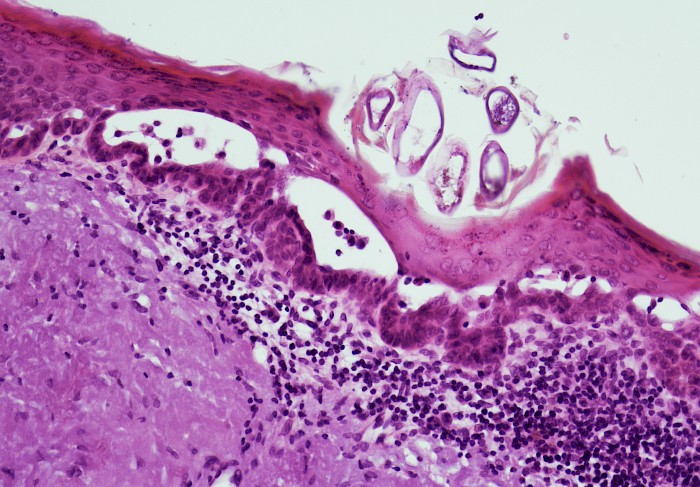
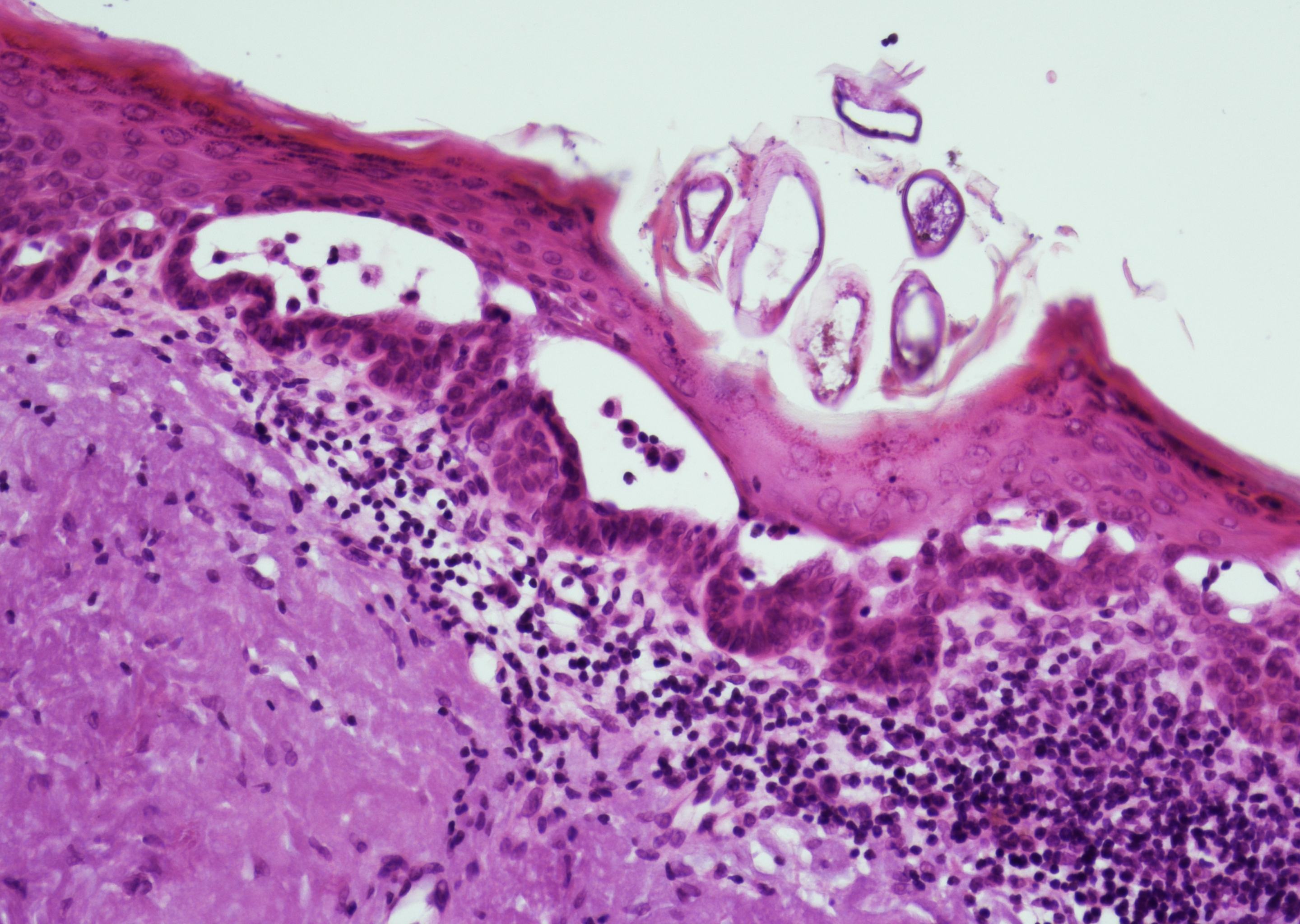 Skin punch biopsy with intracorneal adult…
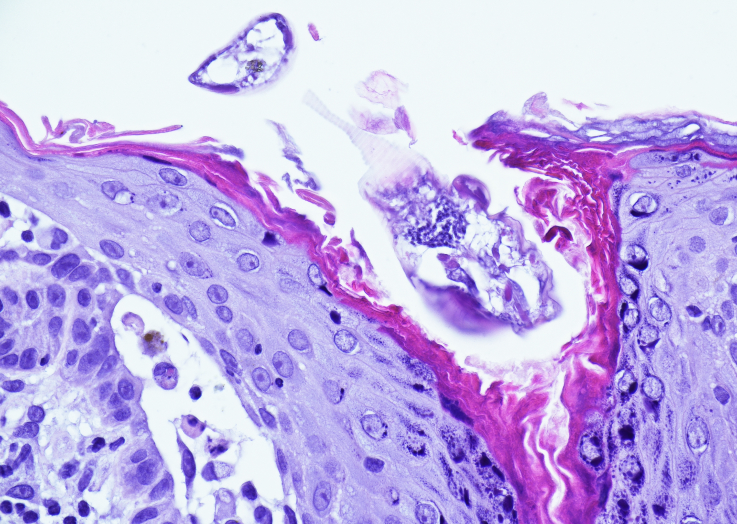 Skin punch biopsy with intracorneal adult…
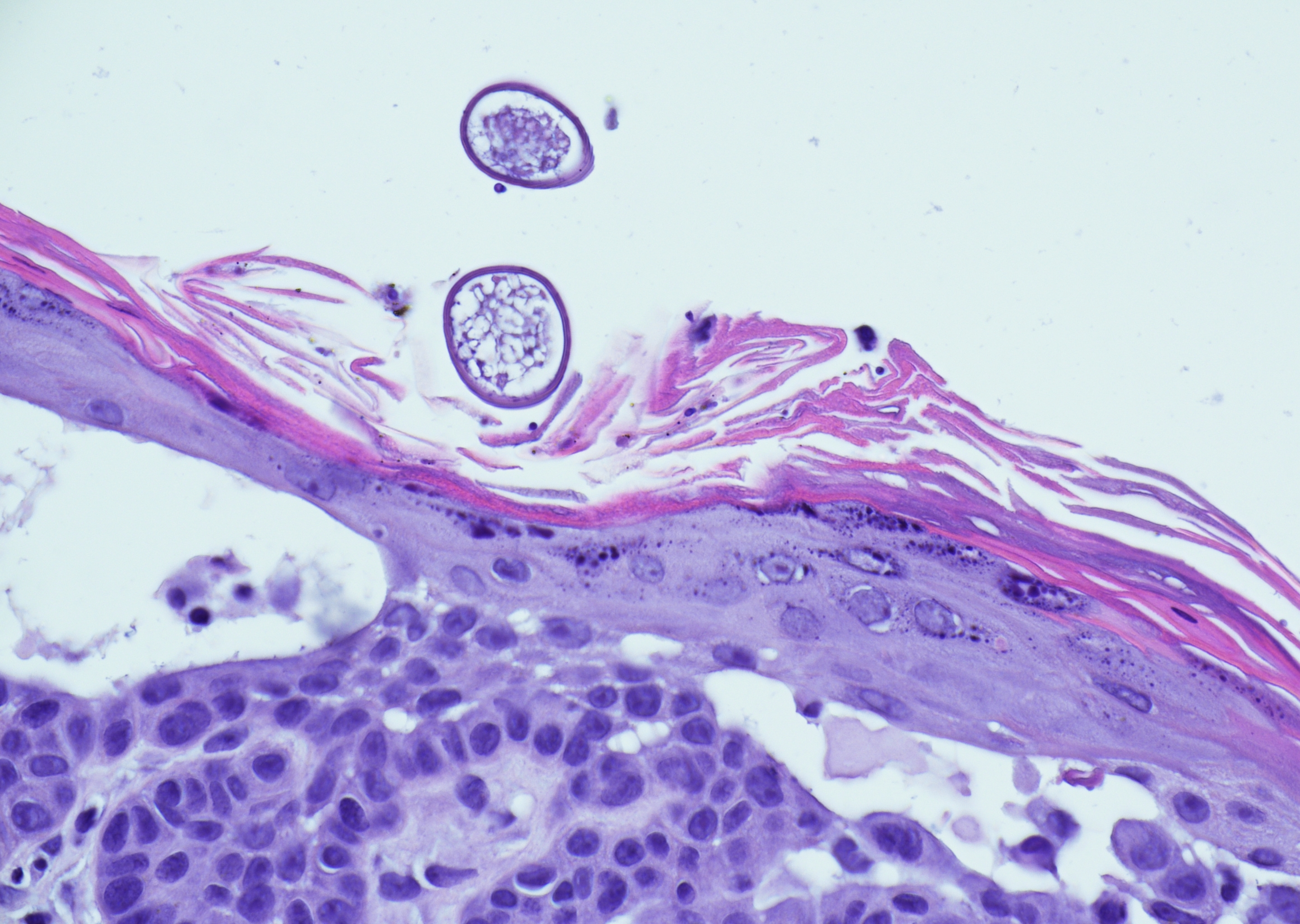 Skin punch biopsy with intracorneal ova and…
Morphology
Histology:
- Skin punch biopsy or shave biopsy with the presence of scabies mites, eggs, or feces (scybala) in the subcorneum or superficial epidermis
- The epidermis may show spongiosis with or without features of excoriation or acantholysis
- Superficial dermal perivascular lymphohistiocytic inflammation with eosinophils and possible neutrophils
- Scabies mites are ovoid (0.5 mm in length) with a chitinous exoskeleton, striated muscles, dorsal spines, and 4 pairs of legs
- Ovoid eggs with 0.1 - 0.2 mm in length
- Mites and their feces show weak birefringence under polarized light
- Histological identification usually is not part of the diagnostic work-up routine, but can be helpful in atypical manifestations
Clinical and morphological manifestations (1):
- Classic scabies
- Papular eruptions with visible burrows and (nightly) pruritus
- Mites typically found at the edge of cutaneous burrows
- Nonspecific inflammatory response in the superficial dermis with eosinophils
- Predilection for palms, interdigital spaces, soles, wrists, axillae, inframammary folds, areola (in women), buttocks, genitals, groins, thighs and umbilicus
- Nodular scabies
- Skin-colored or reddish-brown subcutaneous nodules, due to exaggerated hypersensitivity reaction or from scratching/rubbing
- More prevalent in infants and young children
- Histologic identification of mites less common
- Dense superficial and deep dermal lymphohistiocytic infiltrate and increased eosinophils
- Predilection for axillae, lower trunk, thighs, groin, and scrotum
- Crusted scabies
- Diffuse epidermal hyperkeratosis, pruritus may be mild or absent
- Associated with high mite load
- Histologic features show psoriasiform hyperplasia with massive epidermal hyper- and parakeratosis, as well as innumerable mites between the keratotic scales, and multilayered burrows
- Predilection for palms, ears, extensor surfaces of the elbows, and soles, but can involve any part of the body
- More prevalent in immunocompromised and debilitated populations
- Secondary bacterial infections common
- Bullous scabies
- Rare variant
- Extremely itchy tense or flaccid bullae, with or without visible classic scabies lesions
- Predilection for trunk and extremities
- More prevalent in elderly patients
- Histologic subepidermal blister formation with intraepidermal neutrophilic and eosinophilic infiltration; mites less commonly found; direct and indirect immunofluorescence tests are negative (DD Bullous pemphigoid) (2)
- Nail scabies
- Mostly seen in crusted scabies in immunocompromised patients
- Discolored, thick, and dystrophic finger- and toenails with possible subungual involvement
Differential diagnosis:
- Demodex mites - associated with hair follicles and lack dorsal spines
- Arthropod bite - dermatitis with eosinophils; older lesions may show diffuse or nodular inflammation
- Cutaneous larva migrans - similar presentation of spongiotic and perivascular chronic dermatitis with numerous eosinophils; rarely, nematode larval forms in deep dermis identified
- Eczematous dermatitis - similar histologic appearance with absence of scabies mites, eggs, and feces
- Bullous pemphigoid vs. bullous scabies
Clinical notes
Acquired skin infestation of Sarcoptes scabiei var. hominis mites.
Worldwide distribution and highly contagious.
Transmission via direct skin-to-skin contact and sexual intercourse, or less frequently, indirectly via fomites, such as bedding or clothing, increasing the risk of transmission through duration and frequency of direct skin-to-skin contact, as well as the number of mites on the skin. Commonly, multiple family members, friends, and intimate contacts can be affected simultaneously.
Clinical symptoms typically arise 4-8 weeks after primary inoculation and include intense generalized pruritus that worsens at night due to increased mite activity.
Scabies in the elderly population often presents atypically, with possible involvement of the scalp and face, as well as an increased risk of developing crusted scabies due to various age-related factors.
Scabies in infants causes increased irritability and failure to thrive and typically presents as papules, vesicles, and pustules, whereas in children it commonly presents with burrows, erythematous papules, and nodules.
Diagnosis is usually established clinically, with importance to the patient's history and clinical presentation.
To prevent transmission and recurrence due to reinfestation, all close contacts must be treated simultaneously, even if they are asymptomatic.
Ancillary Testing
Dermoscopy; adhesive tape test; skin scraping with microscopy; skin biopsy; optical coherence tomographyReferences
1. Al-Dabbagh J, Younis R, Ismail N. The currently available diagnostic tools and treatments of scabies and scabies variants: An updated narrative review Medicine. 2023;102(21):e33805. doi:10.1097/MD.0000000000033805
2. Su WJ, Fang S, Chen AJ, Shan K. A case of crusted scabies combined with bullous scabies. Exp Ther Med. 2015;10(4):1533-1535. doi:10.3892/etm.2015.2668